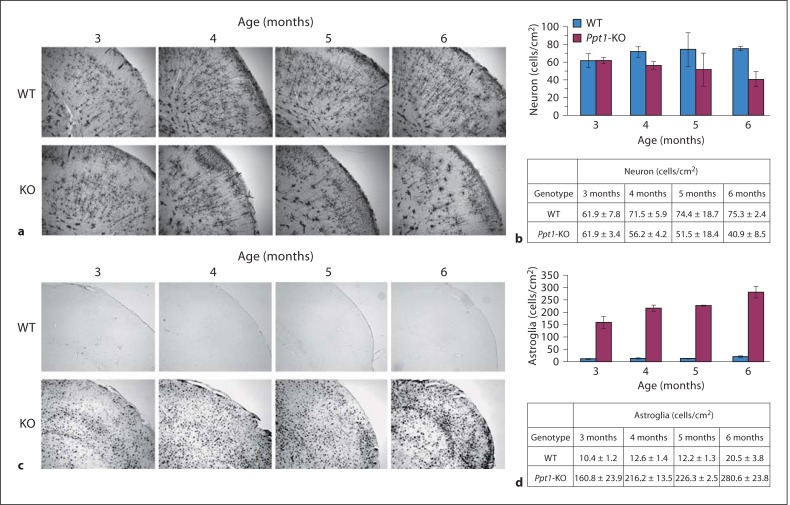
Fig. 4.
Immunohistopathology of brain sections from Ppt1-KO mice and WT littermates. Numbers of neurons and levels of glial fibrillary acidic protein in the brains of Ppt1-KO and WT littermates were determined. a Detection of neuronal cells in P pt1 -KO and WT littermates. Cortical neurons from Ppt1-KO and WT mice (3-, 4-, 5- and 6-month-old) were visualized by the Golgi- Cox staining. b Quantification of the neurons. Three different areas of the cortical tissue were picked randomly, and the cell bodies were countered. Stand deviation was calculated and Student's t test was used to determine the significant difference. c Immunohistochemical detection of astroglial cells in Ppt1-KO and WT littermates. d Quantitation of the astroglial cells. Three different areas of the cortical tissue were randomly chosen to determine the number of astroglia/mm2 area. The results are expressed as the means of 3 determinations ± SD.

