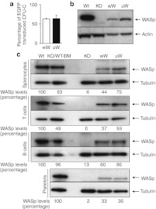
Figure 3.
Expression of human Wiskott–Aldrich syndrome protein (WASp) in hematopoietic cells after transduction. (a) Proportion of colony-forming unit in culture (CFU-C) colonies positive for the integrated foamy virus (FV) vector. Transduction efficiency in Lin-/Sca-1 cells was assessed by real-time-PCR before transplantation. Means ± SD of three independent experiments are shown. (b) WASp expression in transduced Lin-/Sca-1 cells. Western blot (WB) analysis was performed 5 days after transduction. (c) WASp expression in spleen cells and platelets from peripheral blood. WB analysis of whole splenocytes and spleen T and B-cells is shown. Platelets from peripheral blood were also analyzed for WASp expression. Results of densitometry analysis of band intensity are also shown.

