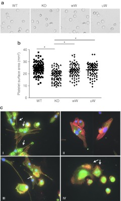
Figure 5.
Improvement of platelet spreading and podosome formation in bone marrow (BM)-derived dendritic cells (DCs) after gene therapy. (a) Morphology of wild-type (WT), KO, and gene therapy- treated (wW, uW) murine platelets. Purified platelets were placed on fibrinogen-coated plate for 45 minutes in the presence of 1 U/ml thrombin and imaged. Results are representative of three experiments in each group. (b) The mean surface area of adherent platelets in WT, KO, and gene therapy-treated mice, was quantified using ImageJ software. *P < 0.05 as compared to KO group, Student t-test. (c) Bone marrow cells, harvested from WT, KO, and gene therapy-treated mice, were cultured in the presence of mGM-CSF and interleukin (IL)-4 for the induction of DCs. Actin is represented in red, vinculin in green and DAPI in blue. (a) Podosome structure in DC from WT mice. (b) Absence of podosome in DC from KO mice. (c,d) Reconstitution of podosomes in gene therapy-treated mice. Arrows indicate podosomes in DCs.

