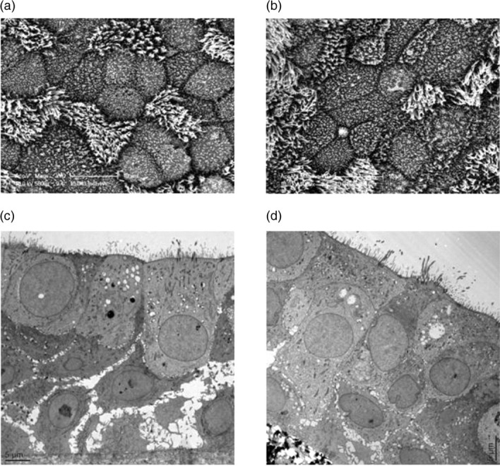
Figure 2.

Paired scanning electron microscopy (SEM) and transmission electron microscopy (TEM) images of EpiAirway tissues with or without NTHi co-culture. One EpiAirway insert was infected with strain R2866 for five days, then fixed and cut in half. One half was processed for SEM (a), the other for TEM (c). Another EpiAirway insert was not infected with NTHi and used as the control for SEM (b) and TEM (d). (a) SEM of infected EpiAirway tissue. (b) SEM of control EpiAirway tissue. (c) TEM of infected EpiAirway tissue. (d) TEM of control EpiAirway tissue. NTHi, non-typeable Haemophilus influenzae
