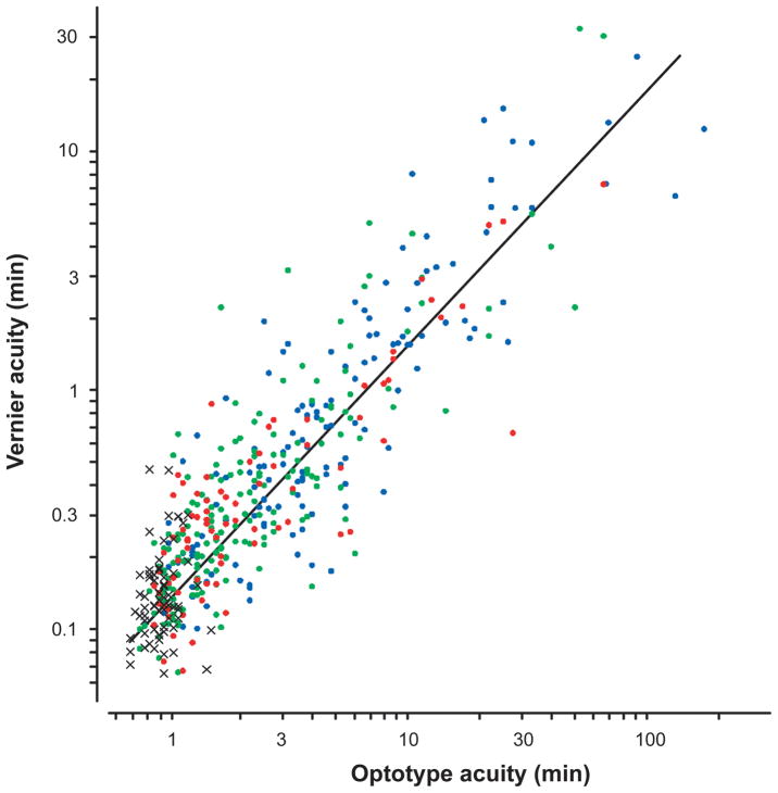
Figure 1.
Vernier acuity vs. Snellen acuity for the non-preferred eye for the entire sample of McKee, Levi and Movshon.17 The crosses show the normal observers. The dots show the amblyopic eyes of aniometropic (green), strabismic (blue) and mixed (i.e., both stabismic and anisometropic – red) subjects.

