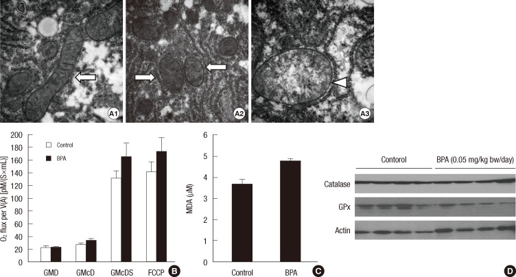
Fig. 5.
Structure and function of hepatic mitochondria in the mice treated with BPA (0.05 mg/kg bw/day) for 5 days. (A) Morphology of hepatic mitochondria by transmission electron microscopy (500,000 × magnification). Control (A1) and BPA-treated mice (A2, A3). Normal mitochondria (arrows) and swollen and cristae-disrupted mitochondria (arrowhead). (B) Oxygen consumption rate of hepatic mitochondria. GMD, glutamate + malate + ADP; GMcD, GMD + cytochrome c; GMcDS, GMcD + succinate; FCCP, uncoupler. (C) MDA concentrations in the liver. (D) Catalase and GPx3 in the liver.

