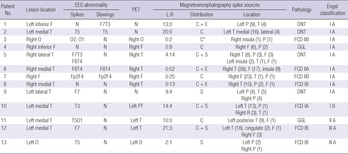
Table 2.
Summary of lesion locations, interictal EEG findings, FDG PET, MEG spike source distribution, pathology, and postsurgical Engel classification of 13 patients
Number in parenthesis shows the number of magnetic spike sources located to the specific brain region. *MEG spike source cluster was located distant from the lesion. L, left; R, right; F, frontal; T, temporal; O, occipital; P, parietal; N, no abnormality; C, cluster; E, extension; S, scatter; DNT, dysembryoplastic neuroepithelial tumor; FCD, focal cortical dysplasia; GGL, ganglioglioma.

