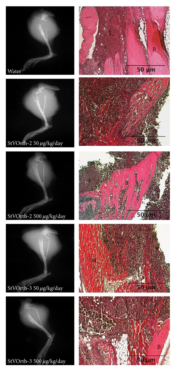
Figure 4.

Tumour invasion. Plain radiographs (left) of tumour-bearing limbs show extensive osteolysis of proximal tibiae and soft tissue extension for all treatment groups. Haematoxylin and eosin-stained sections of orthotopic tumour (right) show tumour cells (T) invading bone (B) and skeletal muscle (M).
