
Figure 1.
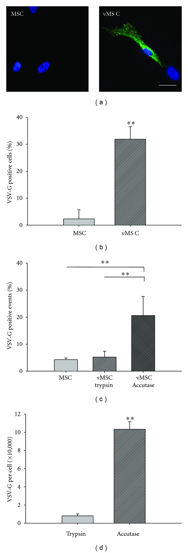
Expression of VSV-G in MSCs. VSV-G expression was analyzed via immunofluorescence using an anti-VSV-G-FITC antibody. (a) Representative image analysis of untransfected MSCs and vMSCs; VSV-G (green); DAPI (blue). Scale bar = 25 μm. (b) Transfection efficiency was defined as the number of VSV-G-positive cells divided by the total number of nuclei and is reported as the mean ± standard deviation. A low level of nonspecific binding was associated with the anti-VSV-G antibody and is reflected in the percentage of positive cells reported in the untransfected population of MSCs (2.3% ± 3.4%). (c) Dissociation reagent impacts VSV-G expression. Trypsin treatment reduces detection of VSV-G expressing cells to that of untransfected MSCs. Accutase treatment retains a significantly greater number of cells expressing VSV-G than treatment with trypsin; **P < 0.005. (d) Dissociation reagent impacts the number of VSV-G proteins per cell. The number of VSV-G proteins expressed per cell is significantly reduced with trypsin treatment as compared with Accutase treatment, which was quantified utilizing Quantum Simply Cellular standards; **P < 0.005.