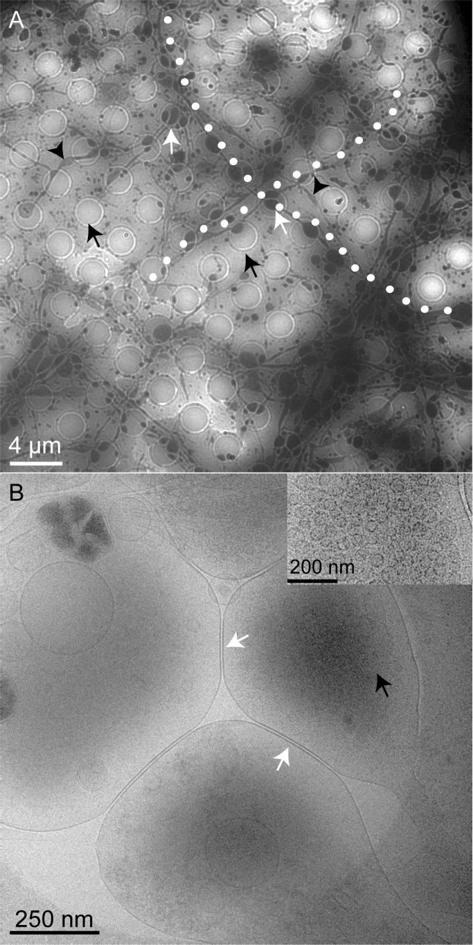
Figure 1.
Representative cryo-EM images showing hippocampal neurons (DIV 8) grown on a sterile, PLL-coated Au/Quantifoil grid. (A) Neuronal processes are visible along the grids, which when on the holes (black arrows) grow a little wider (white arrows) than on the support portion of the grid. The black arrowheads indicate the areas where the axons grow slightly wider outside the holes. Two axonal processes are highlighted (white dots) to indicate the large length scales they are grown on the grid. (B) Magnified view of one of the holes where synapses (white arrows) from closely associated neural processes are visible. High electron density is observed at the areas where synaptic vesicles have accumulated (black arrows). The inset in (B) shows synaptic vesicles observed at the presynaptic boutons.

