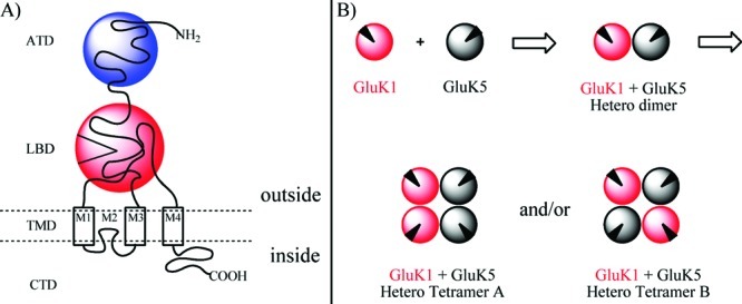
Figure 1.

(A) Cartoon illustration of an iGluR showing the four regions: the carboxylate terminal domain (CTD), the transmembrane domain (TMD), the ligand binding domain (LBD), and the amino terminal domain (ATD). (B) Cartoon illustration of uniform assembly (hetero tetramer A) or opposite assembly (hetero tetramer B) of two heterodimers of GluK1 and GluK5. Black triangle illustrates the LBD.
