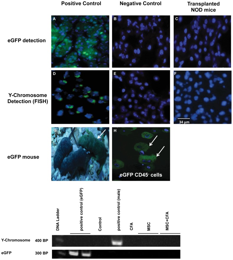
Figure 8. Absence of eGFP and male MSC (CD45−/TER119−) donor cells in salivary tissues of female NOD mice.
Top panel: eGFP detection of MSC donor cells by immunostaining. (A) Salivary tissues from the eGFP donor mouse (positive control) versus (B) the non-eGFP mouse (negative control). (C) NOD mice transplanted with MSCs were negative for eGFP cells. Middle panel: Y-chromosome detection of male MSCs by FISH. (D) male and (E) female salivary tissues used as positive and negative controls, respectively. (F) Female NOD mice transplanted with male MSCs showed no Y-chromosome signal in their salivary glands. Bottom panel: (G) CByB6F1-eGFP transgenic mouse (arrow) and (H) the isolated donor eGFP MSCs before transplantation. Cell nuclei are stained in blue (Hoechst 33258). (I) PCR amplification did not detect the Y-chromosome and eGFP signal in salivary glands of MSCs transplanted NOD mice (n = 11).

