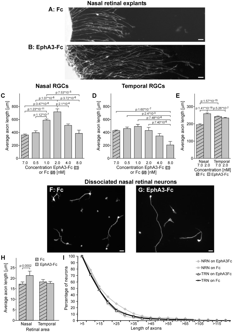
Figure 3. EphA3 ectodomain stimulates nasal RGC axon growth in vitro.
(A, B) Microphotographs of nasal retinal explants grown on clustered Fc (A) or clustered EphA3-Fc at 2 nM (B). RGC axons grow longer on EphA3-Fc. Scale bars = 20 µm. (C–D) Quantification of axon length of nasal (C) and temporal explants (D) grown on substrates formed by laminin and clustered Fc or EphA3-Fc at different concentrations. Axon length is indicated in µm and concentrations are indicated in nM of Fc or EphA3-Fc. Temporal explants grow longer axons than nasal ones in control conditions (p: 0.0002, compare first bar of nasal RGCs in C with first bar of temporal RGCs in D). EphA3-Fc increased nasal RGCs axon growth from 1 to 4 nM showing a peak at 2 nM. Temporal RGCs did not present any significant change in axon growth on EphA3-Fc between 0.5 and 4 nM and presented a significant decrease at 8 nM (ANOVA and Tukey postest, 3 independent experiments, n: 20 longer axons for explant, 3 explants for condition). (E) Quantification of axon length of nasal and temporal explants exposed to soluble clustered Fc or EphA3-Fc at 2 nM. Nasal RGC axons grow significantly longer with EphA3-Fc (ANOVA and Tukey postest, 3 independent experiments, n: 50 longer axons for explant, 3 explants for condition). (F–G) Dissociated nasal retinal neurons immunolabeled against neuron specific βIII tubulin. They present longer axons on clustered EphA3-Fc at 2 nM (G) than on clustered Fc (F). Scale bars = 10 µm. (H) Quantification of axon length of nasal and temporal dissociated retinal neurons grown on clustered Fc or EphA3-Fc at 2 nM. Nasal retinal neurons grow significantly longer axons on EphA3-Fc. (ANOVA and Tukey postest, 3 independent experiments, n: nasal retinal neurons on EphA3-Fc: 288, nasal retinal neurons on Fc: 328, temporal retinal neurons on EphA3-Fc: 636, temporal retinal neurons on Fc: 646). (I) The plot depicts the distribution of axon length of nasal and temporal dissociated retinal neurons (NRN and TRN) grown on EphA3-Fc versus Fc. Values given on the y-axis indicate the proportion of retinal neurons which axons reach the length shown on the x-axis. Nasal retinal neurons present a higher proportion of axons between 20 and 40 µm (n: nasal retinal neurons on EphA3-Fc: 288, nasal retinal neurons on Fc: 328, temporal retinal neurons on EphA3-Fc: 636, temporal retinal neurons on Fc: 646). Results are shown as mean +/– SE.

