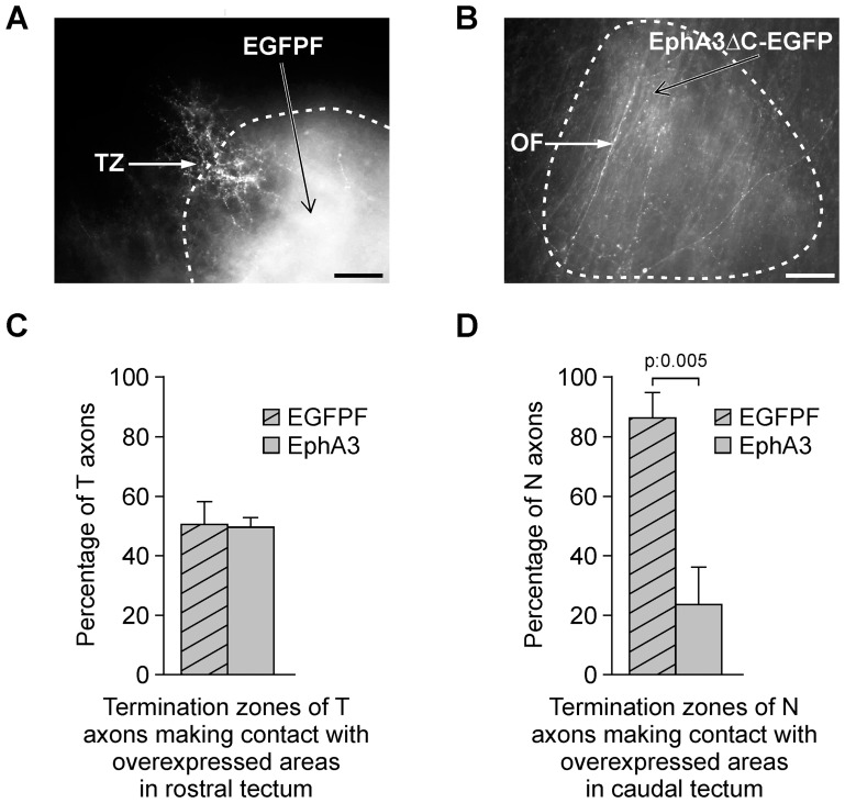
Figure 6. EphA3 ectodomain overexpression stimulates nasal optic fibers (OFs) passing throughout and inhibits termination zones (TZs) formation.
After infection of the optic tectum at E2 with RCAS-BP-B-EGFPF (control) (A) or with RCAS-BP-B-EphA3ΔC-EGFPN3 (B) and DiI labeling of the naso-dorsal retina at E16 (HH42), the tectum was analyzed in whole mounts at E18 (HH44). (A, B) Microphotographs show a termination zone (TZ) formed by nasal optic fibers in an EGFPF-positive domain located in the caudal tectum (A) and nasal optic fibers (OFs) passing throughout an EphA3ΔC-EGFP- positive domain located in the caudal tectum (B). Dotted lines demarcate the overexpressed regions. Scale bars = 50 µm. (C, D) Comparison between the proportions of temporal (C) and (D) nasal RGC axons (T RGC and N RGC) which form TZs in the areas which express EGFPF versus EphA3ΔC-EGFP. Temporal RGC axons were evaluated in the rostral tectum whereas nasal RGC axons were evaluated in the caudal tectum. (C) No significant difference is detected between the proportion of temporal RGC axons which form TZs in EphA3ΔC-EGFP-positive regions and in EGFPF-positive regions (control) in rostral tectum. (D) A significantly lower proportion of nasal axons form TZs in EphA3ΔC-EGFP-positive regions than in EGFPF-positive regions (control) in caudal tectum (Student's t test, n: 4 EphA3ΔC-EGFP-overexpressed tecta versus 7 control tecta for temporal RGCs; n: 5 EphA3ΔC-EGFP-overexpressed tecta versus 4 control tecta for nasal RGCs). Results are shown as mean +/− SE.

