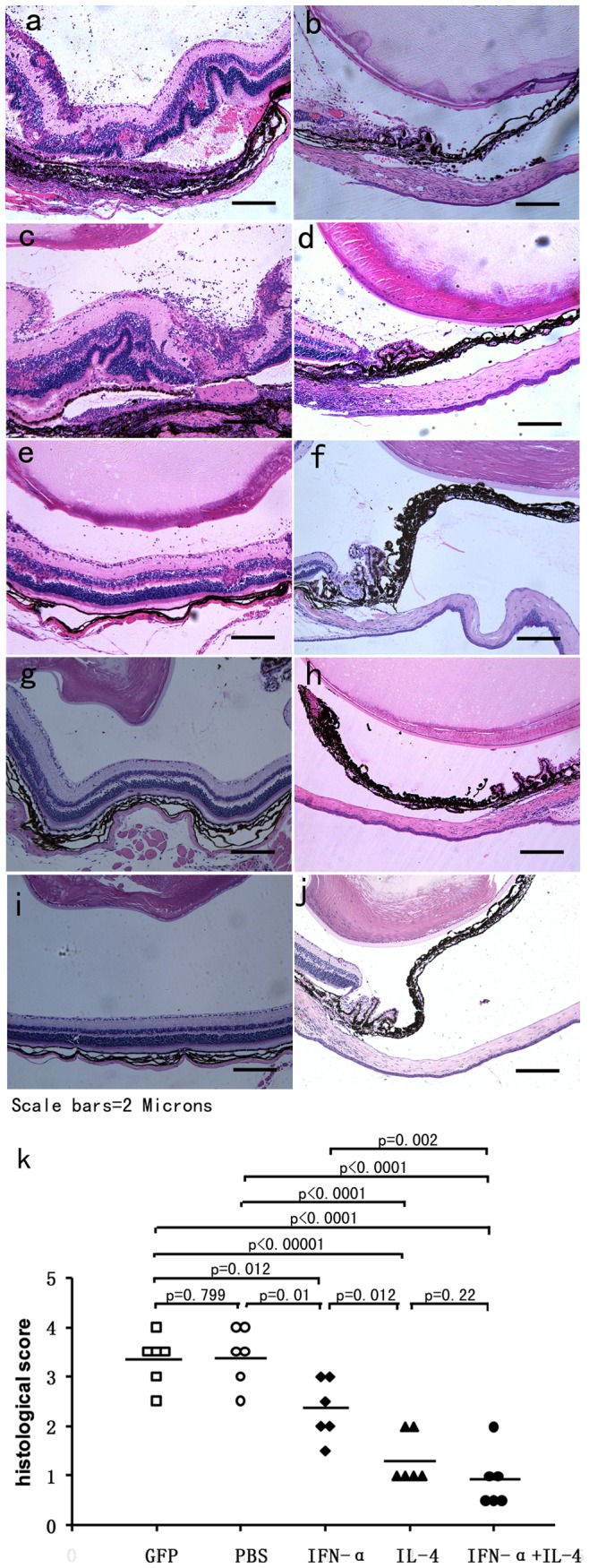
Figure 4. Histological examinations on day 14 of EAU.

Images of histological analysis show severe intraocular inflammation in PBS (a,b) and AAV2.GFP injected eyes (c,d) compared with AAV2.IFN-α treated (e,f), AAV2.IL-4 treated (g,h), and AAV2.IFN-α combined with AAV2.IL-4 treated eyes (i,j). (haematoxylin eosin staining, original magnification×100). EAU was significantly reduced in AAV2.IL-4 treated group, AAV2.IL-4 combined with AAV2.IFN-α treated group as compared with controls (k) (p<0.0001, Mann-Whitney U test). The AAV2.IFN-α treated group also shows a significantly decreased uveitis (p = 0.005). Each point is the score of an individual eye. The mean scores of each group are denoted by the horizontal bars.
