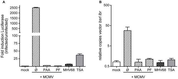
Figure 3. pEpibo-luc-ori is amplified upon MCMV infection.
luc-ori cl.1 cells were infected with MCMV (white hatched bars), MHV68 (black hatched bars) at an MOI of 1 or left untreated (white bar, mock), or treated with 330 nM TSA. In addition, the DNA replication inhibitors PAA (300 µg/ml) and PF (100 µg/ml) were added to infected cells. (A) Bioluminescence assay was performed to determine the FL induction and (B) quantitative realtime PCR was performed 36 h p.i. to determine copy numbers of pEpibo-luc-ori vectors by a PCR specific for the bsr coding sequence compared to the cellular gene lamin B receptor (lbr).

