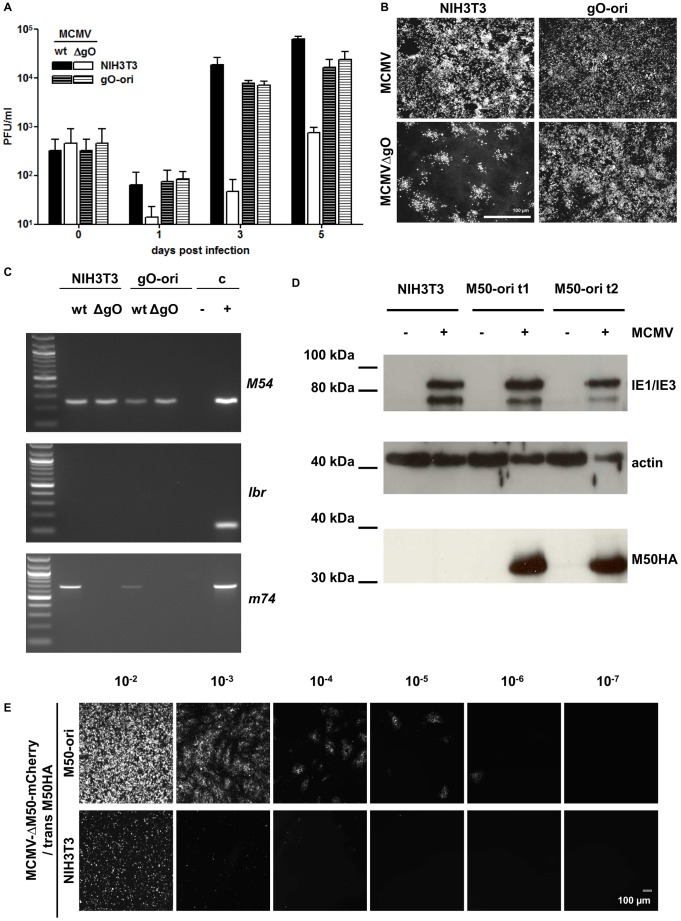
Figure 4. Replicon vector based trans-complementation.
A–C trans-complementation of a glycoprotein. (A) Trans-complementation of the late glycoprotein gO can be facilitated by oriLyt-induced gene expression. NIH3T3 (striped bars) or gO-ori (plain bars) cell lines have been infected with MCMV-wt (white bars) or MCMVΔgO (black bars) at an MOI = 0.05 and centrifugal enhancement. At the indicated time points the number of the infectious virus was quantified in the culture supernatants by standard plaque assay. (B) Immunofluorescence microscopy of infected NIH3T3 or gO-ori cells performed 5 days post infection (MOI = 0.05). Infected cells were stained with the anti-IE antibody CHROMA-101. While MCMVΔgO is restricted to focal cell-to-cell spread in NIH3T3 fibroblasts, it spreads like wild type in the trans-complementing cell line gO-ori. (C) PCR analysis of virus progeny produced on gO-ori cell line. Viral DNA was isolated from supernatants of the viral growth curve of (A) from day 5, cleared from residual cells, and analyzed by PCR. The virus polymerase gene, M54 served as a positive control for viral infection. The cellular gene lbr, served as a control for the lack of residual genomic DNA. PCR on the m74 gene, encoding gO, showed the presence of the gene in MCMV-wt and the lack of it in MCMVΔgO. No uptake and recombination of gO after passage over gO-ori cells could be detected. D–E Trans-complementation of M50, a protein essential for nuclear export of viral capsids. (D) Detection of the M50HA protein (∼35 kDa) in cell lysates of NIH3T3, M50-ori t1 and M50-ori t2. The respective cell lines were infected with MCMV-wt at an MOI of 1 and cell lysates harvested 36 h p.i. (E) Growth of MCMVΔM50-cherry on complementing and non-complementing cell line. Supernatant of the reconstitution of MCMVΔM50-cherry on M50-ori cl.2.1 was serial diluted and used to infect NIH3T3 or M50-ori cells. The trans-complemented virus MCMVΔM50-cherry/M50HA could spread in M50-ori cells, but produced only primary infection in NIH3T3.

