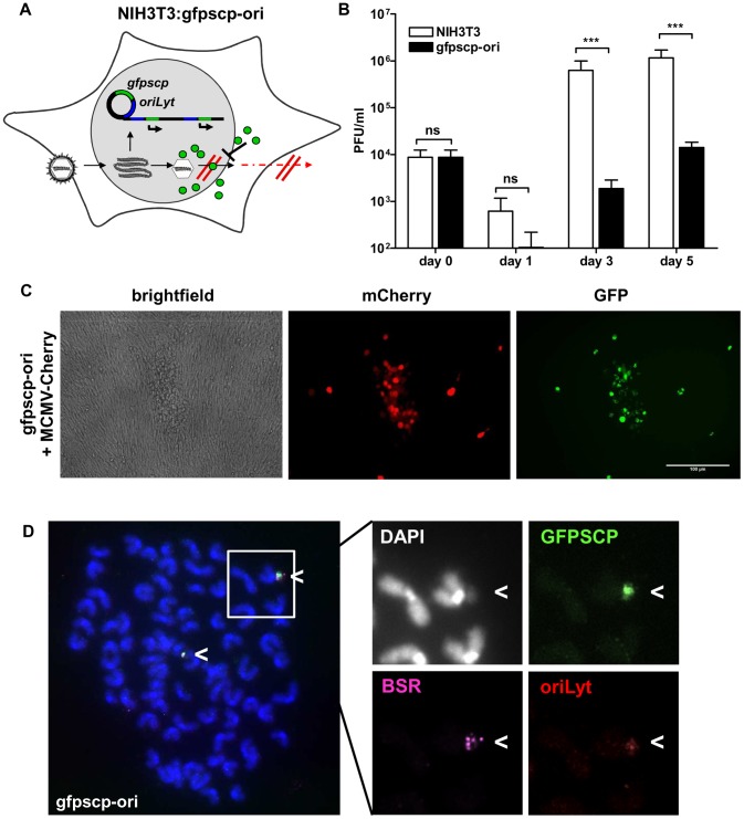
Figure 5. Induction of a dominant-negative protein by the replicon vector inhibits viral growth.
(A) In the transgenic cell line NIH3T3: gfpscp-ori (gfpscp-ori), infection with MCMV induces replication of the construct and thereby activates the production of the dominant-negative protein GFPSCP (green symbols). This protein blocks egress from the nucleus and viral spread is inhibited. (B) NIH3T3 (white bars) and gfpscp-ori (black bars) cell lines were infected with MCMV-wt at MOI of 0.1. At the indicated time points, infectious virus was quantified in the supernatants by standard plaque assay. Due to the production of the inhibitory protein, the cell line gfpscp-ori releases 100–200 - fold fewer viruses into supernatants. (C) NIH3T3 or gfpscp-ori cell lines were infected with MCMV-mCherry at an MOI of 0.1 and expression of GFPSCP and mCherry was assessed 5 days p.i. by fluorescence microscopy. The mCherry protein is expressed with late kinetics and serves as an infection marker. Only in infected cells GFPSCP is produced and localizes according to its typical pattern in nuclear speckles. Plaques on the gfpscp-ori cell line are reduced in size. (D) FISH of metaphase spreads of uninfected gfpscp-ori cells (4n = 76). Three different probes complementary to the gfpscp gene (green), bsr gene (pink) and oriLyt (red) were used. Probes co-localized to DAPI stained extrachromosomal spots, indicating an episomal persistence of pEpibo-gfpscp-ori.

