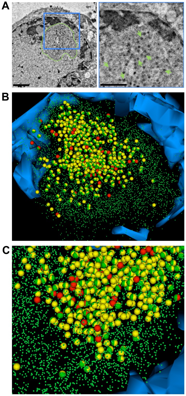
Figure 6. PML protein is associated with entrapped VZV capsids inside PML cages.
Melanoma cells that express doxycycline-induced PML IV were infected with VZV for 48 hours and then high pressure frozen, freeze-substituted, embedded in LR-White resin and labeled with anti-PML polyconal rabbit antibody and Protein A conjugated with 15 nm gold particles. (A) A representative TEM image from a series of seven consecutive 100 nm sections is shown. The area in the blue square (left panel) is shown at higher magnification in the right panel. PML specific gold labeling (green arrows) identifies the PML cage (surrounded by a green line, left panel) in the nucleus. Scale bars are 500 nm. (B) The 3D model shows the electron dense heterochromatin (blue) and the location of mature capsids (63; red spheres), immature capsids (403; yellow spheres) and all PML-specific gold particles (5,219; small green spheres) that were identified in the serial sections. Entrapped mature capsids with associated PML labeling are shown as red/green spheres and immature capsids with PML labeling are shown as yellow/green spheres. (C) Same 3D model as in B but at higher magnification and in a different angle. See also Video S9.

