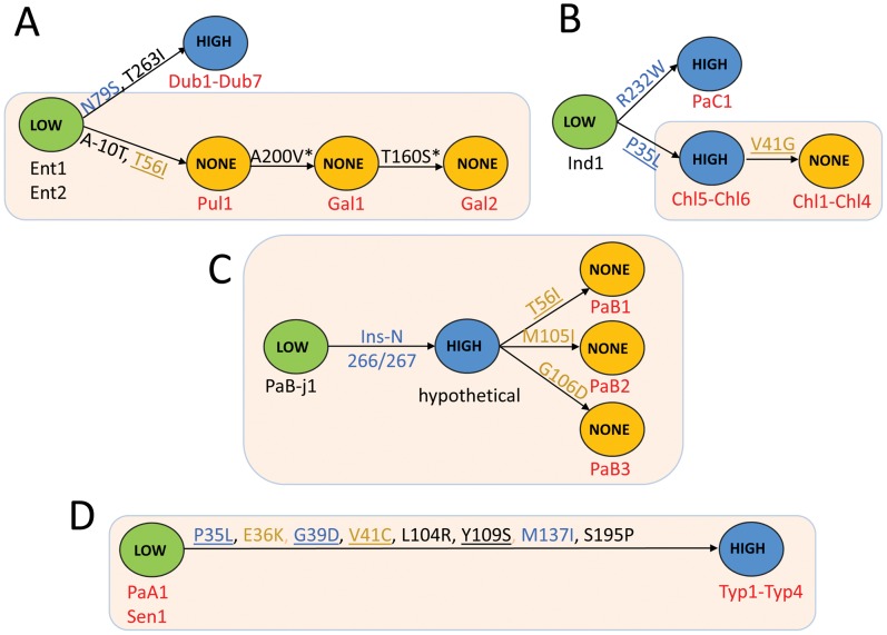
Figure 5. Schematic representation of evolutionary changes in the FimH binding phenotype of selected S. enterica serovars.
The evolutionary changes in the FimH of S. Enteritidis, S. Pullorum, S. Gallinarum and S. Dublin (A), S. Indiana, S. Paratyphi C and S. Choleraesuis (B), S. Paratyphi B (C) and S. Paratyphi A, S. Sendai and S. Typhi (D). Low (green)-FimH with low-binding phenotype; High (blue)-FimH with high-binding phenotype; None (orange)-inactive variant of FimH. The strain tags of systemically non-invasive serovars are in black and the systemically invasive serovars in red. Structural mutations are given along each arrow. Structural hot-spot mutations are underlined. The activating mutations are in blue and the inactivating mutations are in orange. The FimH variants from strains with phylogenetic relatedness supported by MLST are shown in tan boxes.

