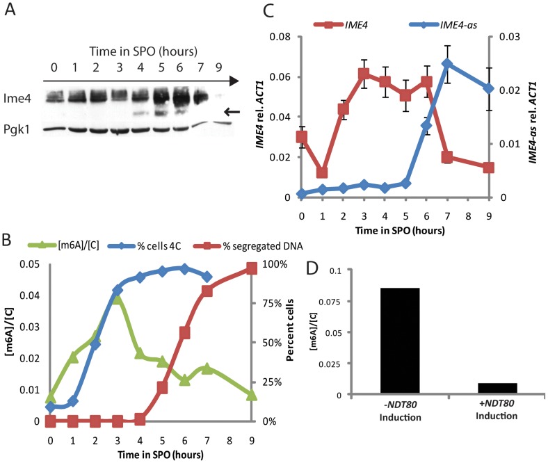
Figure 4. m6A accumulates prior to meiotic divisions.
A) Western analysis for 3x-myc-tagged Ime4 protein (SAy914) throughout meiosis; Pgk1 protein serves as loading control. B) Quantification of m6A abundance relative to cytosine throughout meiosis (green triangles, left axis) in a wild-type strain (SAy821). Percent of 4C cells as quantified by FACS (3×10⁴ cells/time point—blue diamonds, right axis) and percent cells undergoing nuclear divisions as assayed by DAPI staining (200 cells/time point—red squares, right axis) are shown as references for meiotic progression. C) Strand-specific qPCR for sense IME4 (red squares, left axis) and antisense transcript (IME4-as) (blue diamonds, right axis) transcript throughout meiosis. D) m6A relative to cytosine quantification in cells carrying an estradiol-inducible NDT80 construct as their sole source of NDT80 (SAy995). Cells were treated with β-estradiol or vehicle 6 hours after meiotic induction and monitored at 9 hours.

