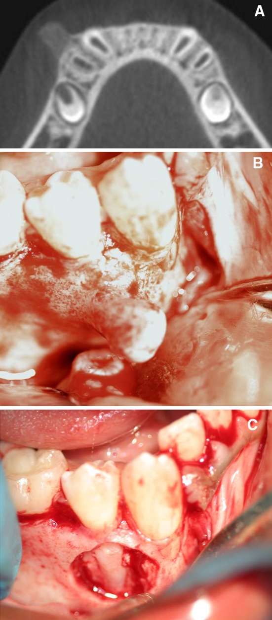
Fig. 3.

a A section from the cone-beam volumetric CT. Note the base of lesion is clearly visible. b The recurrent lesion resembles a similar bony protuberance as seen before. c The recurrent lesion was removed in addition to the associated cortical bone. Note the exposed roots of the canine and premolar
