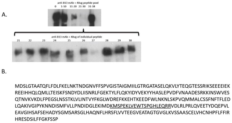
Figure 7. Epitope mapping of anti-serpinB13 mAb.

(A) Western blot analysis of serpinB13 staining with a mAb raised against this protein and preincubated with sequential pools of peptides (upper) or individual peptides (lower) corresponding to serpinB13. A 40 μg pool of peptides or individual peptides were incubated with anti-serpinB13 mAb in a volume of 100 μL at 4°C overnight and used for staining. (B) Full AA sequence of serpinB13. The sequence corresponding to peptide 27, which blocks staining with anti-serpinB13 mAb, is underlined.
