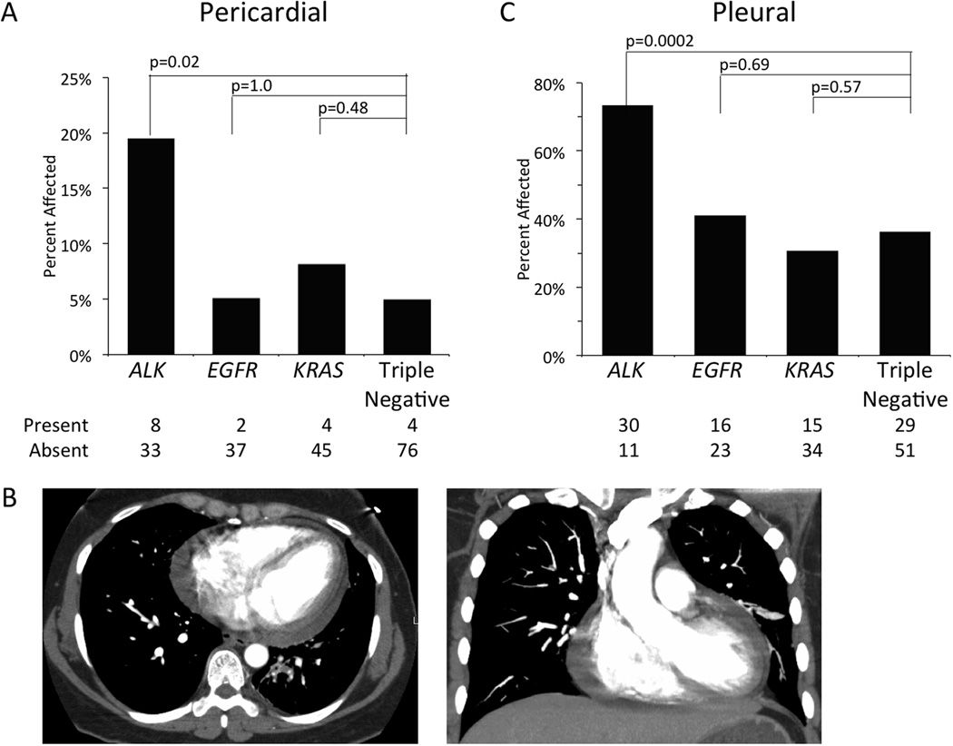
Figure 1. Metastases to Serous Membranes.
The percentage of patients with the presence of metastatic disease in the pericardium (A) or pleura (C) is shown by molecular cohort. Absolute numbers of patients with or without metastases in each cohort is provided below. Panel B depicts axial (left) or coronal (right) CT scan slices showing a pericardial effusion in a 42 year old female patient with an ALK gene rearrangement.

