FIGURE 6.
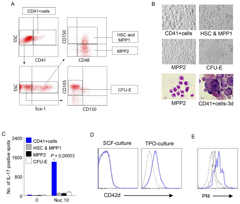
c-Kit+CD41+cells are the Th17 inducers in the Lin−c-Kit+CX3CR1− cell subset. (A) Isolated Lin−c-Kit+CX3CR1− cells were further sorted into CD41+cells, HSC (Sca-1+CD150+CD48−) and MPP1 (Sca-1+CD150+CD48+), MPP2 (Sca-1+CD150−CD48+), and CFU-E (Sca-1−CD150− FcγRIII/II− CD105+low/−). (B) c-Kit+CD41+cells differentiated into megakaryocytes in 3 day culture with IL-3, TPO, EPO and SCF (right bottom panel). Phase contrast of each subset on 3 day culture (top and middle panels) and Giemsa staining of MPP2 cells (left bottom. Bar = 10μm) and cultured CD41+cells (right bottom. Bar = 20μm). Culture conditions for determining hematopoietic lineage potential are in Methods. (C) Sorted c-Kit+CD41+cells (from A) exclusively induced Th17 response to nucleosomes when co-cultured with SNF1 T cells. The sorted subsets of Lin−c-Kit+CX3CR1− cells are designated by color key, and Th17 responses are shown by bars. Mean ± s.e.m. of 5 independent experiments. (D–E) Lin−c-Kit+pure cells differentiate into CD42+cells and produce platelet related molecule, Pf4. FACS shows Lin−c-Kit+pure cells differentiated into CD42+ cells in the presence of TPO but not in SCF alone (D). Nucleosomes feeding increased the production of Pf4 in Lin−c-Kit+pure cells (intracellular staining). PBS (black line) = 14.0% Pf4+ cells vs. Nucleosome (blue) = 55.4% Pf4+ cells in c-Kit+ (CD117+) cell gated population (E). Dotted line = isotype control.

