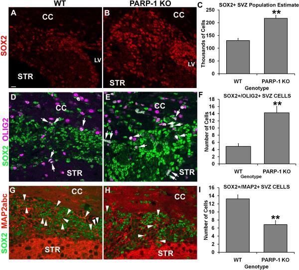
Figure 1. PARP-1 deletion enhances Sox2/Olig2+ precursor cells in the SVZ.
Immunofluorescence labeling was performed to identify Sox2, Olig2, Map2abc and DAPI positive cells. PARP-1 KO mice (B) appear to have more Sox2-positive cells in the SVZ than their WT counterparts (A). To confirm this, we performed unbiased stereology on the SVZ to obtain a population estimate for this region. Stereological analyses confirmed that there was a significant increase in the number of Sox2-positive cells in the SVZ of PARP-1 KO mice compared with WT controls (C, **p<0.01). To confirm the progenitor fate of these cells, we performed immunofluorescence labeling for Sox2, Olig2, Map2abc and DAPI. Confocal microscope images were collected and analyzed to determine the number of Sox2+/Olig2+ or Sox2+/Map2abc+ double-labeled cells within the SVZ. We found more Sox2+/Olig2+ cells present in the SVZ of PARP-1 KO mice (E) than in WT mice (D). Quantification confirmed a significant increase in these cells in the PARP-1 KO mice compared with WT mice (F, **p<0.01). In addition, PARP-1 KO mice appeared to have fewer Sox2+/Map2abc+ double-labeled cells present in the SVZ (H) than their WT counterparts (G). These observations were confirmed with quantification (I, **p<0.01). All data are shown with SEM. Scale bar: 15 μm; CC: corpus callosum; LV: lateral ventricle; STR: striatum.

