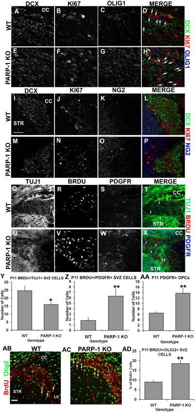
Figure 3. PARP-1 deletion shifts SVZ neural stem cells from neuronal to glial fate.
Mice were injected with BrdU 2 hours prior to sacrifice. Immunofluorescence labeling was performed with Olig1, NG2 or PDGFR to identify OPCs, BrdU or KI67 to identify proliferating cells, and DCX or TUJ1 to identify neuroblasts. More KI67+/Olig1+ double-labeled cells appeared in the SVZ of PARP-1 KO mice (F, G, arrows in H) than in WT mice (B, C, arrows in D). Numerous DCX+ cells were present in WT (A) and PARP-1 KO (E) mice, however, more KI67+/DCX+ cells appeared in the WT SVZ (D) than in the PARP-1 KO SVZ (H). Triple label immunofluorescence with DCX/KI67/NG2 further revealed a greater presence of DCX+/KI67+ in WT mice (I, L) than in PARP-1 KO mice (M, P). We observed an increase in NG2+ cell presence in the SVZ of PARP-1 KO mice (O) along with an increased presence of NG2+/KI67+ SVZ cells (P) compared to WT mice (K, L). To further identify cell fate changes, we examined TUJ1/BrdU/PDGFR triple labeling in the SVZ. We observed a strong presence of TUJ1 in the WT (Q) and PARP-1 KO mouse (U), with many BrdU+/TUJ1+ cells also present in the WT (T) and PARP-1 KO (X) SVZ. More PDGFR+ cells appeared in the SVZ of PARP-1 KO mice (W) than in the WT (S, arrows in T) with an enhanced presence of BrdU+/PDGRF+ cells in the SVZ of PARP-1 KO mice (arrowheads in X) compared to WT mice (T). We quantified the number of BrdU+/TUJ1+ and BrdU+/PDGFR+ cells in the SVZ. We found significantly fewer BrdU+/TUJ1+ cells in the SVZ of PARP-1 KO mice compared with WT mice (Y, *p<0.05). In addition, we found significantly more BrdU+/PDGFR+ cells in the SVZ of PARP-1 KO mice than WT mice (Z, **p<0.01). We quantified PDGFR1+ cells in the SVZ to see if the total number of OPCs varied between WT and KO and we found significantly more PDGFR1+ cells in the SVZ of PARP-1 KO mice than in WT mice (AA, **p<0.01) We also performed immunofluorescence staining with antibodies to BrdU and Olig2 to identify and further quantify proliferating OPCs in the SVZ. The number of BrdU+ and BrdU+/Olig2+ cells was quantified using confocal microscopy. PARP-1 KO mice (AC) appear to have more BrdU+/Olig2+ cells (arrows) present in the SVZ than their WT counterparts (AB). Quantification revealed that PARP-1 KO mice have almost twice as many BrdU+/Olig2+ cells in the SVZ as their WT counterparts (AD, **p<0.01). Scale bars: 10 μm in A for A–H; 50 μm in I for I–X; 25 μm in AB for AB–AC; CC: corpus callosum; STR: striatum; LV: lateral ventricle.

