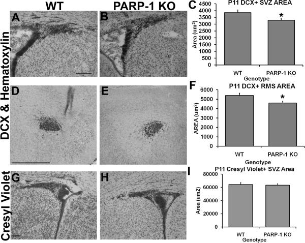
Figure 4. PARP-1 deletion decreases the presence of DCX+ SVZ and RMS area without altering overall SVZ size.
Sections were immunostained with an antibody to DCX to identify the neuroblasts (type A cells) in the SVZ and RMS. The DCX+ area of the dorsolateral SVZ and RMS was obtained using Image J software. We found decreased DCX+ SVZ area in PARP-1 KO mice (B, C, *p<0.05) and this was significantly reduced compared to WT mice (A). We also observed a decrease in the area of DCX+ cells in the RMS of PARP-1 KO mice (E) compared with WT mice (D), which was significant upon quantification with Image J (F, *p<0.05). There was no difference in the total SVZ area as measured with Image J on cresyl violet stained sections in WT (G) or PARP-1 KO mice (H–I). All data are shown with SEM. Scale bars: 125 μm in A for A–B; 500 μm in D for D–E; 100 μm in G for G–H.

