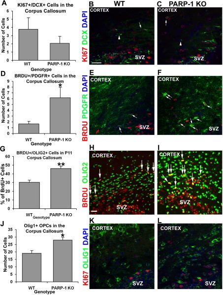
Figure 5. Loss of PARP-1 enhances oligodendrocyte progenitor production in the corpus callosum.
Mice were injected with BrdU 2 hours prior to sacrifice. We performed immunofluorescence staining with antibodies to BrdU or KI67 and PDGFR, Olig1, and Olig2 to identify proliferating OPCS and with DCX to identify neuroblasts. We found no difference in the number of KI67+/DCX+ neuroblasts in the corpus callosum (A) between P11 WT (A, arrow in B) and PARP-1 KO mice (A, arrow in C). The number of BrdU+/PDGFR+ cells significantly increased in the corpus callosum of PARP-1 KO mice (D, *p<0.05, arrows in F) compared with WT mice (D, arrows in E). To confirm increased OPC proliferation, we counted BrdU+/Olig2+ cells in the corpus callosum. We observed a significantly greater percentage of BrdU+ cells co-expressing Olig2 in the corpus callosum of PARP-1 KO mice (G, **p<0.01, arrows in I) than in WT mice (G, arrows in H). In addition, we counted the number of Olig1+ cells present in the corpus callosum. We found significantly more Olig1+ OPCs in the corpus callosum of PARP-1 KO mice (J, *p<0.05, L) than in WT mice (J–K). Some Olig1+ cells co-labeled with KI67, both in WT mice (K) and PARP-1 KO mice (L). All data are shown with SEM. Scale bars: 25 μm in B for B–F, K–L; 25 μm in H for H–I; SVZ: subventricular zone.

