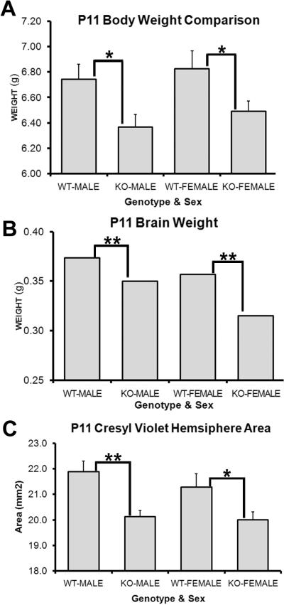
Figure 7. PARP-1 deletion decreases postnatal brain and body size.
Mice were weighed at P11 and then sacrificed for wet brain weight. (A) Both male and female PARP-1 KO mice weighed significantly less than their WT counterparts (*p<0.05). (B) The entire brain was removed and weighed immediately after sacrifice. PARP-1 KOs had significantly smaller brains than male and female WT mice (**p<0.01). To confirm that this change was a result of differences in forebrain size, rather than cerebellar or hind-brain changes, brains were sectioned and stained for cresyl violet. Hemisphere areas were measured on 4 sections per animal at the level of the SVZ. (C) PARP-1 KO mice had significantly smaller forebrain hemispheres than WT mice, with slightly larger difference between males (p<0.01) than females (p<0.05). All data are shown with SEM.

