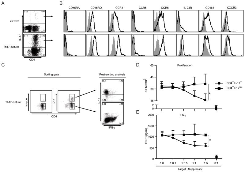
Figure 2. Isolation, Expansion and Characterization of CCR4+CCR6+Th17 cells.
A- Frequency of IL-17 secreting CD4+ T cells in ex vivo PBMCs or in vitro cultured CD4+ memory T cells. Cells were stimulated with PMA/Ionomycin and stained for CD4 and intracellular IL-17. Result shown is representative of 6 independent experiments.
B- Characterization of CD4+IL-17+ T cells. FACS analysis of ex vivo isolated (top) and in vitro primed Th17 cells (bottom) for different surface markers (black lines) and isotype controls (filled histograms). Shown are representative histograms of 4 independent experiments.
C- Sorting of Th17 cells based on CD4 and IL-17 expression and post sorting analysis. In vitro primed Th17 cells were stimulated and sorted as described in Materials and Methods. CD4+IL-17+ and CD4+IL−17− cells were collected as shown. The sorted cell populations were restimulated with PMA/Ionomycin to determine IL-17 and IFN-γ secretion in parallel. Shown is representative dot plot of 3 independent experiments.
D and E- Sorted CD4+IL-17+Th17 or CD4+IL17neg T cells were γ-irradiated and co-cultured with autologous CD3+CD8+ T cells as described in Materials and Methods. Proliferation (D) and IFN-γ secretion (E) of CD8+ T cells are shown as the cumulative result of 3 independent experiments. *p<0.05.

