FIGURE 2.
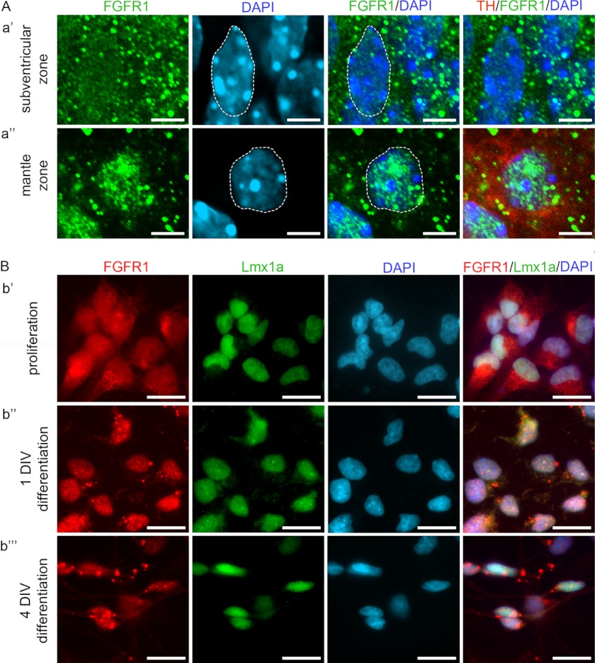
Nuclear accumulation of FGFR1 during differentiation of VM cells. A, confocal microscope images showing subcellular localization of FGFR1 in cells of the mDA field in VM of E14.5 mouse embryos. In the subventricular zone (panel a′), where undifferentiated cells are located, FGFR1 (green) showed a nearly uniform distribution in the cytoplasm and nucleus (blue, dotted line). In the mantle zone (panel a″), FGFR1 (green) shows a prominent accumulation in the nuclear (blue, dotted line) proportion of the differentiated neurons expressing TH (red). Scale bar, 5 μm. B, epifluorescence microscope images showing the accumulation of FGFR1 during differentiation of primary neuronal cultures of rat ventral mesencephalic progenitor cells. During proliferation (panel b′) FGFR1 (red) was localized in the cytoplasm and nucleus (DAPI, blue) of Lmx1a-positive (green) progenitor cells. After differentiation for 1 DIV (panel b″), FGFR1 showed a mainly nuclear distribution in Lmx1a-positive cells. After 4 DIV in differentiation medium (b⁗), FGFR1 was accumulated in the nucleus and also present in the neurites of the neuron like-shaped cells expressing Lmx1a. Scale bar, 10 μm.

