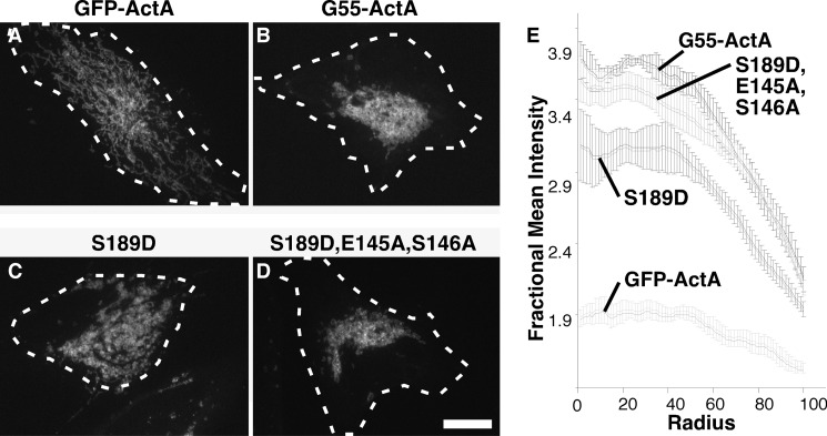
FIGURE 5.
Phosphomimic mutation disrupts membrane tethering. A–D, HeLa cells transfected with the indicated GFP-tagged, mitochondrially targeted constructs were imaged. Thirty min prior to imaging, the cells were treated with brefeldin A to disperse Golgi membranes to ensure that clustering was independent of hypothetical interactions with a centrally positioned Golgi apparatus. Dashed white lines approximate the positions of the cell boundaries. Scale bar = 10 μm. E, the graphs record mitochondrial clustering induced by each construct as measured using the ImageJ radial profile plugin, in which the fluorescence intensity starting from the fluorescence centroid is plotted as a function of radial distance in the first 100 radials (mean ± S.E., n = 4, >15 cells/experiment).

