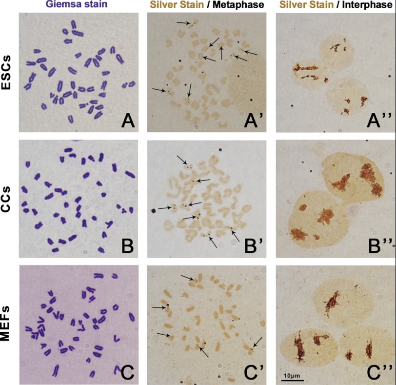
FIGURE 1.
Karyotype and active NORs observation of mouse ESCs, CCs, and MEFs. The metaphase chromosome spreads were stained with 4% Giemsa for karyotyping or stained with silver stain medium to show active NORs. All the three cell lines had normal karyotypes (A, B, and C). The active NORs were stained by AgNO3 and are shown as brown spot pairs on certain chromosomes (arrows). ESCs had more spot pair numbers than CCs and MEFs at metaphase (A′, B′, C′). At interphase, the silver stain signal showed as big brown clusters in the nuclei in all the three cell lines (A′′, B′′, C′′). Bar = 10 μm.

