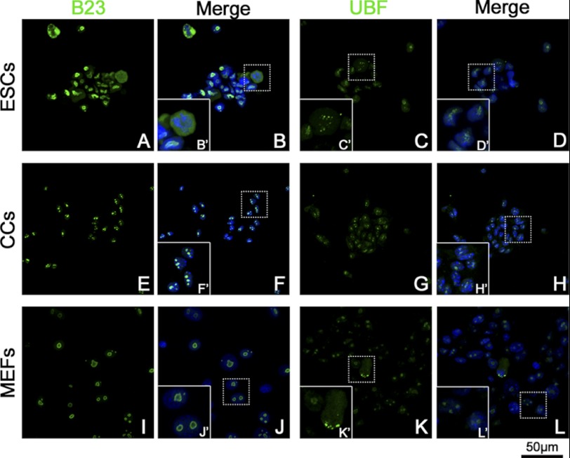
FIGURE 3.
Distribution of B23 and UBF in ESCs, CCs, and MEFs. B23 and UBF were labeled by monoclonal anti-B23 antibody/monoclonal anti-UBF antibody, respectively, and fluorescent conjunct second antibody (green). The nuclei/chromosomes were stained with Hoechst 33342 (blue). Typical areas (dotted line rectangle) were magnified to show the details (solid line rectangle). B23 showed as three or four condensed clusters within the nucleus in CCs (F′). In MEFs, B23 signals showed as 1–3 big rings (J′). ESCs showed much a stronger B23 signal with an irregular shape (A). At metaphase, the B23 signal distributed to the whole cytoplasm (B′). The UBF signal showed as several clusters in the nuclei of all the three groups of cells (C, G, and K). During mitosis, the UBF signal showed as pairs of small spots in metaphase plate (C′) or spindle polar (K′).

