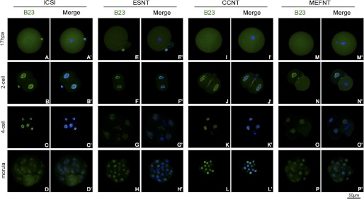
FIGURE 7.
Distribution of B23 in NT and ICSI embryos at difference preimplantation developmental stages. B23s were labeled by monoclonal anti-B23 antibody and fluorescent conjunct second antibody (green). The nuclei/chromosomes were stained with Hoechst 33342 (blue). The B23 distributed to the whole cytoplasm at metaphase of the first mitosis (A, E, I, and M) then recruited in the nuclei at the 2-cell stage (B, F, J, and N). When the blastomere started to cleavage, B23 distributed to the cytoplasm again (N). All the four groups of embryos showed ring-like B23 signals around the nucleoli at 4-cell and morula stages (C, D, G, H, K, L, O, and P).

