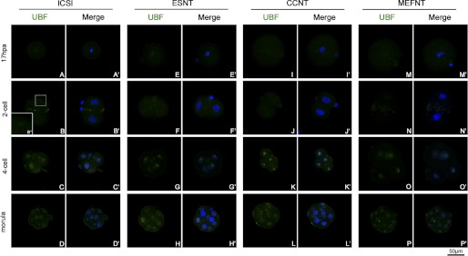
FIGURE 8.
Distribution of UBF in NT and ICSI embryos at difference preimplantation developmental stages. UBFs were labeled by monoclonal anti-UBF antibody and fluorescent conjunct second antibody (green). The nuclei/chromosomes were stained with Hoechst 33342 (blue). No UBF signal could be detected at metaphase of the first mitosis (A, E, I, and M). At the 2-cell stage, no UBF signal could be detected in ESNT, CCNT, and MEFNT embryos (B, F, J, and N). But several ICSI embryos showed weak UBF spots around the nucleoli (B′′). At the 4-cell stage, the UBF signals showed as discontinuous small clusters around nucleoli (C, G, K, and O). At the morula stage, many blastomeres were under mitosis, and the UBF signals distributed in the whole cytoplasm with chromosomes or at the spindle polar (D, H, L, and P).

