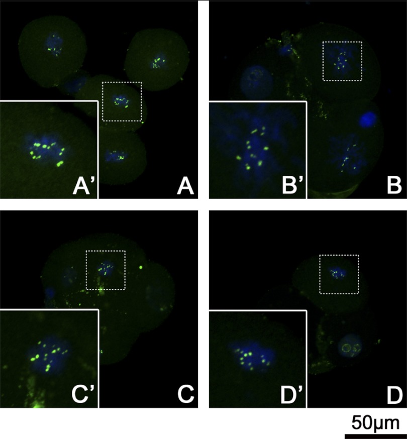
FIGURE 9.
UBF signals of 4-cell stage embryos at metaphase. ICSI embryos (A), ESNT embryos (B), CCNT embryos (C), and MEFNT embryos (D) at 48 h post-activation/sperm injection were blocked with 3 μg/ml nocodazole for 6 h to synchronize to the metaphase. UBF were labeled by monoclonal anti-UBF antibody and fluorescent conjunct second antibody (green). The chromosomes were stained with Hoechst 33342 (blue). Metaphase plate (dotted line rectangle) was magnified to show the details (solid line rectangle). The UBF signals showed as spot pairs on certain chromosomes. Four groups shared the same pattern but with different spot pair numbers.

