FIGURE 7.
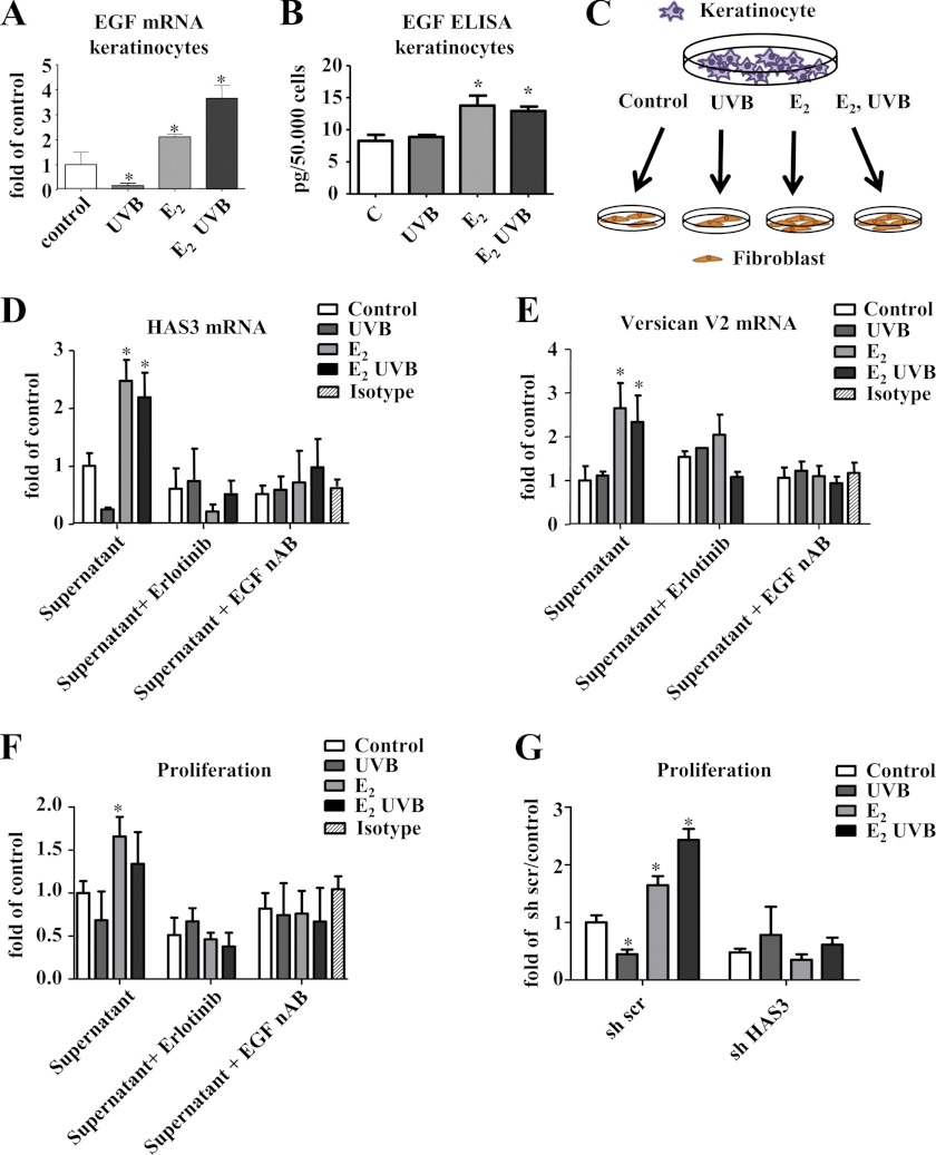
EGF release from keratinocytes in response to E2 and UVB. A, keratinocytes were treated with 100 nm E2, 100 mJ/cm2 UVB, or both. After 24 h, EGF mRNA expression was analyzed by real time RT-PCR. B, EGF protein was determined by ELISA in cell culture supernatants of keratinocytes treated as described in A. C, schematic representation of the experiments shown in D–G using keratinocyte-conditioned medium to investigate paracrine EGF effects. D and E, supernatants as shown in B were used to stimulate human skin fibroblasts. After 24 h, HAS3 mRNA expression and VERSICAN V2 mRNA expression were determined by real time RT-PCR in the presence or absence of 3 μm erlotinib, EGF neutralizing antibody (EGF nAB, 0.35 μg/ml), or isotype control IgG (0.35 μg/ml). F, cell culture supernatants as in B were used to stimulate fibroblasts. [3H]Thymidine incorporation was determined as a measure of DNA synthesis and proliferation; G, [3H]thymidine incorporation in response to the supernatants as in B in fibroblasts pretreated with scrambled or HAS3-targeting lentiviral shRNA; n = 3–6, mean ± S.E.; *, p < 0.05 versus control.

