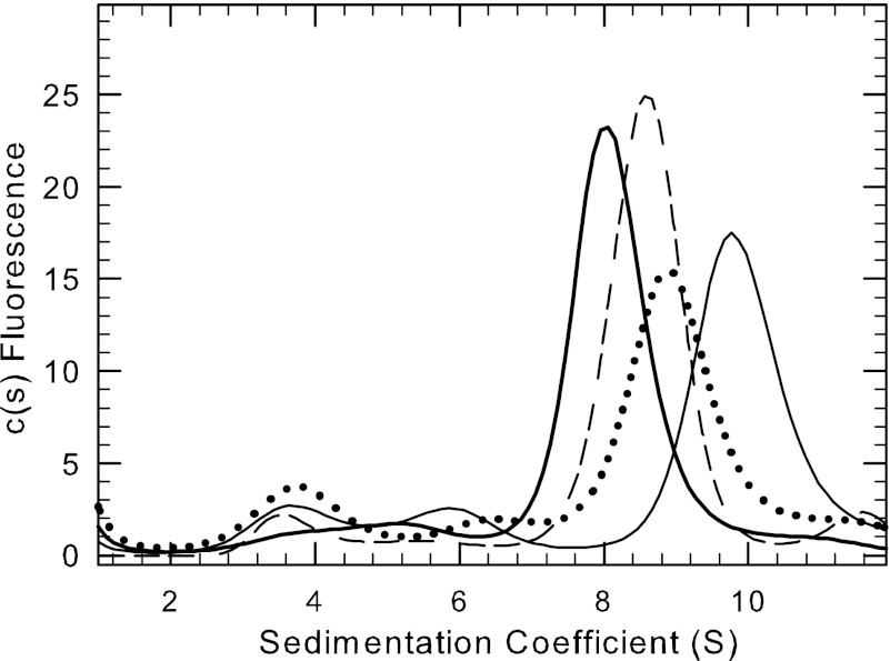
FIGURE 6.
Binding of the G-protein, transducin, to PDE6 holoenzyme. Purified PDE6 holoenzyme (2.5 or 6 nm) covalently labeled with IAF was incubated with various concentrations of purified, activated Tα-GTPγS. Sedimentation velocity analysis was then carried out (50,000 rpm at 20 °C), and the data were analyzed to determine the sedimentation coefficient for each concentration: no added Tα-GTPγS (thick solid line), 3000-fold molar excess of Tα-GTPγS (dashed line), 5000-fold excess (dotted line), and 10,000-fold excess (thin solid line). The traces shown were normalized based on the total protein fluorescence in each sample.

