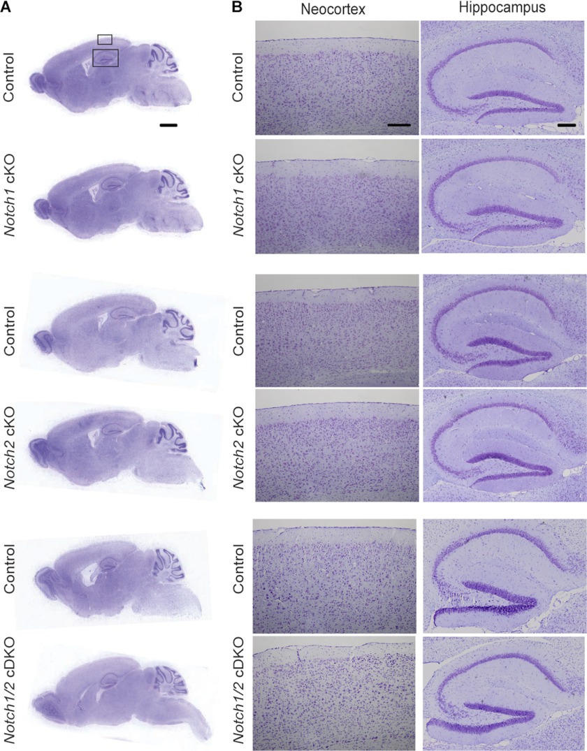
FIGURE 1.
Normal gross morphology of the cerebral cortex of Notch cKO mice. A and B, shown is Nissl staining of comparable sagittal brain sections (10 μm, paraffin) of aged Notch cKO and littermate control mice (24 months for Notch1 cKO, 24 months for Notch2 cKO, 12 months for Notch1/2 cDKO). Representative images of sagittal brain sections at low magnification (A) and the boxed areas of the neocortex and hippocampus at a higher magnification (B) are shown. Scale bar, 1 mm (A) and 200 μm (B).

