FIGURE 7.
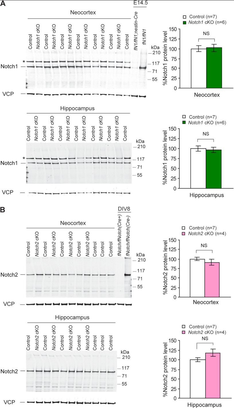
Unchanged levels of Notch1 and Notch2 proteins in the cerebral cortex of Notch cKO mice. A, shown are Western analysis (left) and quantification (right) of Notch1 proteins in the neocortex and the hippocampus of adult Notch1 cKO (n = 6) and littermate control (n = 7) mice using a Notch1 rabbit monoclonal antibody (clone D1E11, Cell Signal Technology). B, shown are Western analysis (left) and quantification (right) of Notch2 proteins in the neocortex and hippocampus of adult Notch2 cKO (n = 4) and control (n = 7) mice using a Notch2 rabbit monoclonal antibody (clone D76A6, Cell Signal Technology). No significant difference of Notch1 or Notch2 proteins was detected between cKO and control samples, indicating that despite the deletion of the floxed exons at the genomic DNA level, protein expression is not altered in cKO cortical samples and suggesting that there is little Notch expression in these excitatory pyramidal neurons of the adult cerebral cortex where Cre is expressed under the control of the αCaMKII promoter. Total protein lysates of E14.5 embryonic brains of Nestin-Cre-driven Notch1 cKO (fNotch1/fNotch1;Nestin-Cre) mice (A) or neuronal cultures (DIV8) derived from fNotch1/fNotch1;fNotch2/fNotch2 (fNotch/fNotch) postnatal pups (B) are included as controls. All values are normalized to that of VCP (Vasolin containing protein) protein, which is used as loading control. The Notch1 antibody (rabbit monoclonal) used here recognizes a nonspecific signal that is only present in the adult brain (*). All data are expressed as the mean ± S.E. Statistical analysis was performed using two-tailed unpaired Student's t test. NS, not significant.

