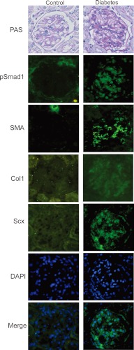
FIGURE 6.
Detection of Scx and SMA in diabetic nephropathy mice. STZ-induced diabetic (right panels) and control mice (left panels) were dissected at 32 weeks after treatment (n = 6 in each group). Light microscopy showed mesangial matrix expansion in STZ mice by periodic acid-Schiff's (PAS) staining. An increase in the expression of pSmad1, SMA, and Scx was noted by immunohistochemical staining in diabetic glomeruli in STZ mice. Nuclear staining of Scx in glomeruli in STZ mice was observed. Nuclei of the cells were stained by DAPI (blue). Representative microscopic appearance of the glomerulus is shown. Original magnification for all panels was ×400.

