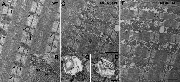
FIGURE 1.
EDL muscle fibers from MCK-βAPP mice showed severe mitochondrial alterations. A and B, in WT EDL fibers from 2–3-month-old animals (n = 3), mitochondria were primarily located in correspondence to the I band, forming two rows on both sides of each Z line (A, arrows). Mitochondria (Mit.) were located in the close proximity of Ca2+ release units or triads (B) and presented a healthy appearance (see “Results” for more detail). C–E, muscle fibers from 2–3-month-old MCK-βAPP mice (n = 3) contained a large number of mitochondria with severely altered morphology (open arrows). These mitochondria were usually swollen and contained vacuoles, myelin figures, or lamellar structures surrounded by an almost completely clear matrix (D and E). F, in some fibers mitochondria were abnormally enlarged and formed columns or clusters between myofibrils and/or under the sarcolemma. Bars, A, C, and F, 1 μm; B, D, and E, 0.2 μm.

