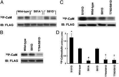
Fig. 3.
[32Pi] biosynthetic labeling of mutant CaM. (A–C) Autoradiographs and immunoblots (IB) of transfected CaM immunoprecipitated from [32Pi] biosynthetically labeled BAEC. BAEC were transfected with WT or mutant CaM cDNA constructs as indicated and biosynthetically labeled with [32Pi]. The lysates were immunoprecipitated with an antibody directed against the FLAG epitope. Immune complexes were analyzed by SDS/PAGE and autoradiography, and membranes were then probed with an anti-FLAG antibody. (D) The results of densitometric analysis of pooled data from the experiments shown in A–C and similar experiments, plotting the fold increase in the 32P-CaM signal, relative to the signal obtained for CaM labeling as analyzed in cells transfected with WT CaM. * indicates P < 0.05, and # indicates P < 0.001 relative to WT CaM-transfected cells; † indicates both P < 0.005 relative to WT CaM-transfected cells and P < 0.01 relative to S81D mutant CaM-transfected cells.

