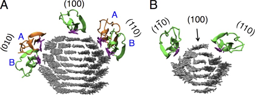
FIGURE 1.
Starting points for MD simulations of the Cel7A CBM1 on the surfaces of cellulose microfibrils. The CBMs are shown in green or orange, whereas the tyrosine residues on the planar face of the CBM are shown in purple. A, the 36-chain microfibril simulations were started with the CBM in the center of the hydrophobic (100) surface or one of two positions on (110) or (010) hydrophilic surfaces. B, for the 16-chain microfibril, simulations were started with the CBM in the center of the (110) and (010) hydrophilic surfaces and on the hydrophobic (100) surface containing a single chain. In all simulations, the microfibril was reoriented such that the z direction is normal to the starting surface, the x direction is along the cellulose fiber axis, and the y direction is perpendicular to the chains in the plane of the starting surface.

