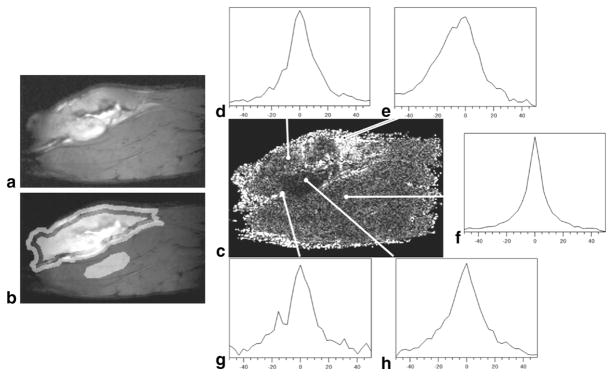
FIG. 1.
a: Water PH image of a tumor-bearing rat leg. b: Water PH image with tumor rim, tumor center, and normal muscle ROIs highlighted. c: Corresponding absolute asymmetry image. Typical spectra observed the (e, g) tumor rim, (d, h) tumor center, and (f) normal muscle. Spectra in the tumor rim are visually more asymmetrically broadened.

