
Figure 1.
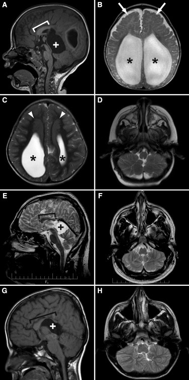
Characteristic Neuroimaging Features of GPSM2-related Chudley-McCullough Syndrome
(A) illustrates posterior agenesis of the corpus callosum (bracket indicates remaining corpus callosum) and a quadrigeminal plate cistern cyst (white plus sign) causing mass effect on the cerebellum and tectum in individual 8A at 3 years of age.
(B) illustrates severe ventriculomegaly (black asterisks) and frontal polymicrogyria (white arrows) in individual 8B at 6 months of age.
(C) illustrates large frontal gray matter heterotopia (white arrowheads) located superior and medial to the enlarged lateral ventricles (black asterisks) in individual 8A at 3 years of age.
(D) illustrates inferior cerebellar hemisphere dysplasia in individual 8B at 6 months of age.
(E) and (F) illustrate a short corpus callosum (bracket indicates remaining corpus callosum) and a quadrigeminal plate cistern cyst (black plus sign) causing mass effect on the cerebellum and tectum as well as cerebellar hemisphere dysplasia in individual 9A (CG6 from Walsh et al.9) at 26 years of age.
(G) and (H) illustrate similar findings in individual 10B (IV-2 from Yariz et al.10) at 12 years of age, although the corpus callosum is thinned posteriorly and dysplastic anteriorly, rather than short.
(A) and (G) are sagittal T1-weighted images; (B)–(D), (F), and (H) are axial T2-weighted images, and (E) is a sagittal T2-weighted image.