
Figure 3.
NCL Pathology in Progranulin-Deficient Mice
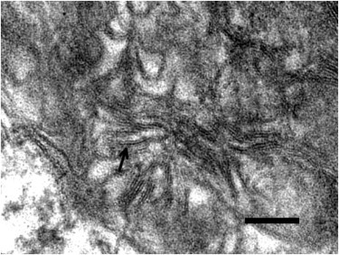
Electron micrograph showing a high-power view of one of the storage granules in a neuron from the habenula of a Grn−/− mouse. The pattern corresponds to the rectilinear complex, which, along with fingerprint profiles, is characteristic of most types of NCL. The arrow points to a pentalaminar profile. The bar indicates 100 nm.