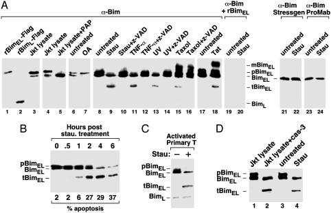
Fig. 1.
Caspase cleavage of pBimEL in T cells at an early stage of apoptosis. (A) Caspase cleavage of pBimEL in apoptotic Jurkat T cells. Anti-Bim Western blotting was performed with the rabbit polyclonal antibodies directed against the full-length BimEL to examine the purified Flag-tagged rBimEL and Flag-tagged rBimL (lanes 1 and 2), Jurkat cell lysates incubated with (lane 5) or without (lanes 3 and 4) PAP, and lysates from Jurkat cells subjected to the various treatments as indicated (lanes 7–20). In lanes 19 and 20, the antibody solution was preincubated with purified rBimEL before being used in Western blotting. In lanes 21–24, Western blotting was performed with anti-Bim antibodies obtained from StressGen Biotechnologies (Victoria, Canada) and ProMab Biotechnologies (Mountain View, CA), respectively. (B) Cleavage of pBimEL at an early stage of apoptosis. Lysates of Jurkat cells treated with staurosporine for the indicated periods of time (hr) were analyzed by anti-Bim Western blotting. The percentage of apoptotic cells was determined by flow-cytometric analysis and is indicated at the bottom. (C) Apoptosis-induced cleavage of pBimEL in activated mouse primary T cells. Anti-Bim Western blot analysis of lysates from activated mouse primary T cells treated with or without staurosporine. (D) pBimEL cleaved in vitro by recombinant caspases-3 comigrates with tBimEL from apoptotic Jurkat cells as indicated by anti-Bim Western blotting. Lanes 1 and 2, lysates from healthy Jurkat cells were incubated with or without recombinant caspase-3. Lanes 3 and 4 show lysates from Jurkat cells treated with or without staurosporine. Stau, staurosporine; OA, okadaic acid; mBimEL, modified BimEL.

